Micosis superficiales: Dermatofitosis (Tiñas)
RESUMEN
En los países desarrollados la mayoría de las infecciones micóticas de la piel son producidas por hongos pertenecientes a los géneros Malassezia, Candida y Dermatofitos. La pitiriasis versicolor y las infecciones cutáneas por cándida son tratadas en otros posts.
En este post se revisa la etiología, epidemiología, diagnóstico y tratamiento de las dermatofitosis o tiñas mas frecuentes atendida por el médico de Atención Primaria, infecciones causadas por dermatofitos, un grupo de hongos capaces de invadir e infectar los tejidos queratinizados, pelo, piel y uñas, gracias a la queratinasa que poseen, tanto del hombre como de algunos animales. Las infecciones que producen son generalmente superficiales y afectan fundamentalmente al estrato córneo de la piel y anejos, variando su expresión clínica dependiendo de la región afectada..
ETIOLOGÍA Y EPIDEMIOLOGÍA
La incidencia y aislamiento de las distintas especies de dermatofitos varía mucho de unas regiones a otras del mundo estando influída por múltiples factores como: edad, sexo, grupo étnico, hidratación, humedad, poder patógeno, resistencia del huésped, fuente de infección , etc.
Los dermatofitos se clasifican en tres géneros: Epidermophyton, Microsporum y Trichophyton, si bien hay reconocidas más de 40 especies, de las que alrededor de una docena son patógenas para el hombre. Clásicamente, la denominación de las infecciones causadas por dermatofitos ha estado relacionada con las localizaciones anatómicas involucradas. En nuestro medio, el agente etiológico más frecuentemente implicado es T. rubrum, seguido de T. mentagrophytes y M. canis; T. rubrum es la especie más frecuente causante de tinea pedis, tinea ungium, tinea cruris y tinea corporis en el mundo. Aunque en la actualidad la incidencia de tinea capitis ha descendido considerablemente en los países desarrollados, se ha observado un incremento de los casos de tinea pedis y onicomicosis. Además, las nuevas migraciones han reintroducido especies antropofílicas (T. tonsurans) en países con escasa prevalencia para esta especie. La distribución del resto de especies varía en función de la localización geográfica.
La etiología de las dermatofitosis según las localizaciones anatómicas mas frecuentemente involucradas es:
- Tinea capitis: Habitualmente es causada por especies de Microsporum y Trichophyton, siendo M. canis la especie mayoritaria. Es poco frecuente en países desarrollados, aunque algunos autores sostienen que es la micosis infantil más frecuente. También son frecuentes T. violaceum, T. tonsurans, T. soudanense y M. audouinii.
- Tinea barbae: Está causada frecuentemente por especies zoofílicas de Trichophyton, sobre todo T. mentagrophytes.
- Tinea corporis: Los principales dermatofitos implicados son T. rubrum, M. canis, T. mentagrophytes y T. tonsurans
- Tinea cruris: Los patógenos causantes más comunes son T. rubrum y E. foccosum
- Tinea manuum: La mayoría de los casos están causados por Trichophyton rubrum
- Tinea pedís: Las especies implicadas más frecuentemente son T. rubrum, T. mentagrophytes var. interdigitale y Epidermophyton floccosum
- Tinea ungium: Así se denomina la invasión de la uña por un dermatofito, mientras que cuando ésta está producida por un hongo no dermatofito se denomina onicomicosis, aunque este último término se emplea para la infección fúngica de las uñas en general. T. rubrum y T. mentagrophytes var. interdigitale son las especies más frecuentemente implicadas.
CLÍNICA
Las manifestaciones clínicas puede variar, dependiendo de la región afectada. El prurito es el síntoma más frecuente. Las lesiones de la piel, en general, se caracterizan por una inflamación que es más grave en los bordes, con eritema, descamación y, ocasionalmente, la formación de ampollas.
Tiña del cuero cabelludo
Querion de Celso: Tiña tonsurante o no inflamatoria, que afecta fundamentalmente a niños y provoca alopecia no cicatricial, es decir reversible. Comienza como una pequeña pápula, que se extiende para formar escamas, irregulares o zonas bien delimitadas de alopecia. Es frecuente la presencia de adenopatías cervicales y occipitales.


Favus: También es posible observar un querion o masa inflamada con un componente pustuloso importante; a esta reacción en general le sigue la cicatrización. Las lesiones supurativas, en general, se observan cuando la infección es causada por dermatofitos zoofílicos.


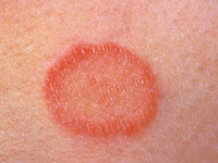
Tiña corporal o herpes circinado
La tiña corporal ocurre en el tronco, las extremidades y el rostro. Se caracteriza por una sola lesión o múltiples lesiones anulares escamosas con un borde eritematoso, escamoso y levemente elevado, márgenes bien definidos y una zona clara en el centro. En los bordes de la lesión se pueden encontrar pápulas, pústulas o vesículas foliculares. Las lesiones son variablemente pruriginosas. En muchas personas, la tiña corporal no tratada se resuelve en varios meses.


Tiña de la barba
La tiña de la barba es una infección del pelo y de la piel de la barba y la zona del bigote y, en general, se observa en los hombres. Las lesiones pueden incluir descamación, pústulas foliculares y eritema. La tiña de la barba se presenta en raras ocasiones, siendo la mayoría de las infecciones cutáneas localizadas en la zona de la barba causadas por bacterias y no por hongos.


Tiña facial
La tiña facial se observa en las partes lampiñas del rostro. Las lesiones son generalmente pruriginosas; empeorando este después de la exposición a la luz solar. Algunas lesiones se parecen a las de la tiña corporal; otras pueden tener muy pocas o ninguna escama o bordes elevados. En algunos casos, las áreas de eritema son indistintas. Debido a la presentación atípica, la tiña facial muchas veces se confunde con otras enfermedades de la piel que afectan a la cara.
Tiña crural o “eccema marginado de Hebra”
Es la infección de las ingles, áreas perianal y perineal y, ocasionalmente, la parte superior de los muslos, frecuente en varones adultos. Las lesiones suelen ser bilaterales y asimétricas y se extienden distalmente desde la cara interna de los muslos. También es conocida como “eccema marginado de Hebra”. Los síntomas incluyen ardor y prurito. Se encuentran pústulas y vesículas en los bordes activos del área infectada, junto con maceración, sobre una lesión de base roja, escamosa y con bordes elevados.


Tiña del pie
Más conocida como “pie de atleta”, afecta al pie, especialmente a la planta y dedos. La forma clínica más frecuente es la intertriginosa con maceración, descamación y formación de fisuras, especialmente en el 4.º espacio interdigital. Otra forma de presentación es la forma hiperqueratósica, con finas escamas grisáceas que cubren la planta, el talón y ambos lados del pie (tiña en mocasín).


Tiña de la mano
La tiña de la mano es una infección dermatofítica que aparece en una mano o, en ocasiones, en ambas manos. En esta afección, las palmas se vuelven levemente secas, escamosas y eritematosas. Con mayor frecuencia, es causada por dermatofitos antropofílicos (estos casos pueden ocurrir como una generalización del pie de atleta), pero en ocasiones puede ser causada por microorganismos zoofílicos.
Tina ungueal
Así se denomina la invasión de la uña por un dermatofito, mientras que cuando ésta está producida por un hongo no dermatofito se denomina onicomicosis, aunque este último término se emplea para la infección fúngica de las uñas en general. La afectación puede ser subungueal (distal y proximal) o superficial (leuconiquia tricofítica). Se caracteriza por uñas engrosadas, descoloridas, rotas y distróficas. La superficie de la uña puede separarse del lecho ungueal.


DIAGNÓSTICO
El diagnóstico de las dermatofitosis es habitualmente clínico, pudiendo confirmarse mediante pruebas complementarias (examen microscópico y cultivo). El examen clínico mediante la Luz de Wood, una luz ultravioleta de longitud de onda larga, puede detectar la fluorescencia en algunos dermatofitos. El examen microscópico con hidróxido de potasio (KOH) puede detectar hifas y conidias en muestras de piel y pelo. Se necesita realizar cultivos micóticos para identificar el microorganismo. También se realizan biopsias de piel y de uñas.
TRATAMIENTO
Tratamiento tópico
La mayoría de las dermatofitosis vistas en Atención Primaria, excepto las del cuero cabelludo y las de las uñas, son leves y a menudo curan con cremas de antimicóticos. Los principios activos de las medicaciones antifúngicas tópicas mas frecuentemente utilizadas son el clotrimazol, miconazol y ketoconazol. Habitualmente, las cremas se aplican dos veces al día y el tratamiento debe prolongarse por lo menos de 7 a 10 días después de que la erupción haya desaparecido por completo. Si se interrumpe la aplicación del tratamiento precozmente, la infección puede no haberse erradicado completamente y reaparecer la erupción. También hay que saber y explicar la paciente que pueden transcurrir varios días antes de que el tratamiento antifúngico tópico surta efecto.
Se debe recomendar, como medida coadyuvante, mantener las zonas infectadas limpias, lavándolas frecuentemente con agua y jabón y secarlas bien a continuación
Si una infección micótica de la piel supura, es posible que también haya podido desarrollarse una infección bacteriana, requiriéndose tratamiento con antibióticos topicos.
Aunque en general, el tratamiento de primera elección de las micosis ungueales es la terbinafina via oral, el tratamiento con antifúngicos tópicos, en forma de lacas, puede ser eficaz en algunas tiñas ungueales, en especial cuando la afectación la parte proximal no se encuentra comprometida (Ciclopirox 8%, 1 aplicación/24 h durante 3-6 meses según se trate de unas de las manos o pies, o amorolfina 5%, 1-2 aplicaciones semana, 6 meses (manos) o 9 meses (pies), limando previamente bordes y superficie ungueal).
Tratamiento sistémico
El tratamiento sistémico es más efectivo que el tópico y se aplicará en los casos de tiña capitis, tiña de la barba, tiña ungueal y otras dermatofitosis con lesiones extensas, hiperqueratósicas, gran componente inflamatorio, etc.
Griseofulvina: tiene una eficacia sobradamente conocida y se ha utilizado como referencia frente a otros antifúngicos, siendo el fármaco de elección en la tiña del cuero cabelludo y de la barba. La dosis normal es de 250-500 mg / 12 horas o 125 mg / 6 horas conjuntamente con las comidas. En niños, normalmente 10 mg / Kg / día en dosis fraccionadas. La duración del tratamiento será de cuatro semanas o en algunos casos hasta la curación clínica de la afección. Entre los efectos secundarios destacan cefalea, trastornos gástricos, fotosensibilidad, erupciones y leucopenia.
Terbinafina: Es el tratamiento de elección en la tiña ungueal, 250 mg 1 vez / día 6 semanas en tiña de las uñas de manos y 12 semanas en tiña de unas de los pies. También es una alternativa al tratamiento con griseofulmina de la tiña capitis, y una de sus principales ventajas es que no inhibe la vía del citocromo p450, a tener en cuenta en pacientes polimedicados.
Itraconazol: Estudios recientes han demostrado que la administración de 200 mg, 1 vez / día durante 7 días o 100 mg 1 vez / día durante 15 días resulta eficaz en el tratamiento de la tinea pedis recalcitrante. También es una alternativa de segunda elección a la terbinafina en la tiña ungueal, 200 mg / 12 h, 1 semana al mes durante 2-3 meses (manos) o 3-4 meses (pies).
Fluconazol: Es un fármaco de segunda elección en el tratamiento de las micosis ungueales, administrado 150 mg/semana durante 3-4 meses respectivamente según sean unas de manos o pies.
Bibliografía recomendada
- Amichai B, Nitzan B, Mosckovitz R, et al. Iontophoretic delivery of terbinafine in onychomycosis: a preliminary study. Br J Dermatol. 2010;162:46-50.
- Baran R, Tosti A, Hartmane I, et al. An innovative water-soluble biopolymer improves efficacy of ciclopirox nail lacquer in the management of onychomycosis. J Eur Acad Dermatol Venereol. 2009;23:773-781.
- Bell-Syer SE, Hart R, Crawford F, et al. A systematic review of oral treatments for fungal infections of the skin of the feet. J Dermatolog Treat. 2001;12:69-74.
- Chang, CH, Young-Xu Y, Kurth T, et al. The safety of oral antifungal treatments for superficial dermatophytosis and onychomycosis: a meta-analysis. Am J Med. 2007;120:791-798.
- Crawford F, Hollis S. Topical treatments for fungal infections of the skin and nails of the foot. Cochrane Database Syst Rev. 2007;(3):CD001434.
- Crawford F, Young P, Godfrey C, et al. Oral treatments for toenail onychomycosis: a systematic review. Arch Dermatol. 2002;138:811-816
- Drake LA, Dinehart SM, Farmer ER, et al. Guidelines of care for superficial mycotic infections of the skin: tinea corporis, tinea cruris, tinea faciei, tinea manuum, and tinea pedis. Guidelines/Outcomes Committee. American Academy of Dermatology. J Am Acad Dermatol. 1996;34:282-286.
- Elewski BE. Treatment of tinea capitis: beyond griseofulvin. J Am Acad Dermatol. 1999;40(6 pt 2):S27-S30.
- Faergemann J, Baran R. Epidemiology, clinical presentation and diagnosis of onychomycosis. Br J Dermatol. 2003;149(suppl 65):1-4.
- Friedlander SF. The optimal therapy for tinea capitis. Pediatr Dermatol. 2000;17:325-326.
- Gonzalez U, Seaton T, Bergus G, et al. Systemic antifungal therapy for tinea capitis in children. Cochrane Database Syst Rev. 2007;(4):CD004685.
- Gupta AK, Adam P, Dlova N, et al. Therapeutic options for the treatment of tinea capitis caused by Trichophyton species: griseofulvin versus the new oral antifungal agents, terbinafine, itraconazole, and fluconazole. Pediatr Dermatol. 2001;18:433-438.
- Gupta AK, Cooper EA, Bowen JE, et al. Meta-analysis: griseofulvin efficacy in the treatment of tinea capitis. J Drugs Dermatol. 2008;7:369-372.
- Gupta AK, Fleckman P, Baran R. Ciclopirox nail lacquer topical solution 8% in the treatment of toenail onychomycosis. J Am Acad Dermatol. 2000;43(4 suppl):S70-S80.
- Gupta AK, Jain HC, Lynde CW, et al. Prevalence and epidemiology of unsuspected onychomycosis in patients visiting dermatologists' offices in Ontario, Canada - a multicenter survey of 2001 patients. Int J Dermatol. 1997;36:783-787.
- Hart R, Bell-Syer SE, Crawford F, et al. Systematic review of topical treatments for fungal infections of the skin and nails of the feet. BMJ. 1999;319:79-82.
- Higgins EM, Fuller LC, Smith CH. Guidelines for the management of tinea capitis. British Association of Dermatologists. Br J Dermatol. 2000;143:53-58.
- Hinojosa JR, Hitchcock K, Rodriguez JE. Clinical inquiries. Which oral antifungal is best for toenail onychomycosis? J Fam Pract. 2007;56:581-582.
- Jahangir M, Hussain I, Ul Hasan M, et al. A double-blind randomized comparative trial of itraconazole versus terbinafine for 2 weeks in tinea capitis. Br J Dermatol. 1998;139:672-674.
- Korting HC, Kiencke P, Nelles S, et al. Comparable efficacy and safety of various topical formulations of terbinafine in tinea pedis irrespective of the treatment regimen: results of a meta-analysis. Am J Clin Dermatol. 2007;8:357-364.
- Macura AB. Dermatophyte infections. Int J Dermatol. 1993:32:313-323.
- Roberts DT, Taylor WD, Boyle J; British Association of Dermatologists. Guidelines for treatment of onychomycosis. Br J Dermatol. 2003;148:402-410.
- Scher RK, Baran R. Onychomycosis in clinical practice: factors contributing to recurrence. Br J Dermatol. 2003;149(Suppl 65):5-9.
- Sigurgeirsson B, Olafsson JH, Steinsson JB, et al. Long-term effectiveness of treatment with terbinafine vs. itraconazole in onychomycosis: a 5-year blinded prospective follow-up study. Arch Dermatol. 2002;138:353-357.
- Tey HL, Tan AS, Chan YC. Meta-analysis of randomized, controlled trials comparing griseofulvin and terbinafine in the treatment of tinea capitis. J Am Acad Dermatol. 2011;64:663-670.
- Trivedi NA, Shah PC. A meta-analysis comparing efficacy of continuous terbinafine with intermittent itraconazole for toenail onychomycosis. Indian J Dermatol. 2010;55:198-199.
- Zhang AY, Camp WL, Elewski BE. Advances in topical and systemic antifungals. Dermatol Clin. 2007;25:165-183.
Te puede interesar:
- Alopecia androgenética: abordaje práctico y novedades terapéuticas
- Corticoides tópicos
- Dermatitis atópica
- Dermatitis seborreica
- Eccema dishidrótico: actualización terapéutica 2025
- Eczema dishidrótico e hiperhidrosis
- Eritema nodoso
- Erupción polimorfa luminica
- Herpes simple
- Herpes Simple en Atención Primaria: Guía 2025 de Diagnóstico,Tratamiento y Prevención
- Herpes zoster
- Liquen plano
- Micosis superficiales: Candidiasis
- Onicomicosis: Guía Actualizada 2025 de Diagnóstico y Tratamiento para Atención Primaria
- Pitiriasis rosada de Gibert
- Pitiriasis Rosada de Gibert (Actualización 2025)
- Pitiriasis versicolor
- Protoporfiria eritropoyética.
- Prurito
- Psoriasis
- Psoriasis: Actualización clínica 2025
- Queratosis actínica
- Reacciones fotoalérgicas a medicamentos
- Sarcoidosis
- Úlceras Orales: Guía Diagnóstica y Terapéutica para Médicos de Familia
- Úlceras de la boca
- Urticaria solar


Disulpe, quería apuntarle lo siguiente:
ResponderEliminarEn contra de lo que usted escribe en su blog; lo que se conoce como Kerion de Celso es una Tinea Capitis INFLAMATORIA.
Sólo hay dos tipos de Tiñas en el cuero cabelludo (Tinea Capitis) no inflamatorias: Microspórica y Tricofítica.
Un saludo
Describiéndolo mas ampliamente el querion de Celso suele comenzar como una tiña capitis no inflamatoria. En ocasiones puede seguirse de una inflamación severa a nivel del folículo piloso, generándose en el área pápulas y pústulas que se secan dejando una costra. Durante este periodo, puede observarse el "signo de la espumadera", que consiste en observar la salida de pus por cada uno de los orificios de los folículos pilosos al hacer presión lateral sobre la zona. El proceso cicatriza de manera fibrosa y deja áreas de alopecia definitiva. A las formas evolucionadas inflmatorias hay algunos autores que los denominan Favus
ResponderEliminar